Included Content
- Organization of the Genome, Replication, Mutation, & Repair
- Regulation of Gene Expression I & II
- Prokaryotic Genetics
- Cell Cycle
- Chromosomal Abnormalities
- Inheritance Patterns & Human Disease I & II
- Epigenetics & Genomic Imprinting
- Skeletal Muscle Physiology I & II
- Smooth Muscle Physiology
- Cardiac Muscle Physiology
- Biochemical Composition of the Skin
- Many DNA-based diagnostic tests use a DNA polymerase from Thermus aquaticusa bacterium that can surface high temperatures. Compared with the DNA of bacteria that grow at 25ºC, the DNA of T. aquaticusis expected to have higher fraction of which of the following nucleotides?
- Adenine and cytosine
- Cytosine and Guanine
- Adenine and Thymine
- Adenine and Guanine
- Cytosine and ThymineAnswer B. Cytosine and Guanine have three hydrogen bonds, as oppose to adenine and thymine which only have two hydrogen bonds.
Genome, Replication, Mutation & Repair.
- Some nitrofurans form adducts with bases in DNA that cannot be repaired via the base-excision repair pathways. The adducts leads to distortions of the DNA double helix. The lesion is most likely repaired by which one of the following DNA repair pathways?
- Homologous recombination repair
- Mismatch repair
- Nonhomologous end joint
- Nucleotide-excision repair
- Base-excision repairAnswer D.
Genome, Replication, Mutation & Repair.
-
A 48-year-old woman had endometrial cancer and undergoes a hysterectomy. Immunohistochemistry of tumor tissue reveals the presence of MLH1 and PMS2, but an absence of MSH2 and MSH6. Based on this finding, the most likely diagnosis is which of the following?
- Cockayne syndrome
- Hereditary breast and ovarian cancer syndrome
- Lynch syndrome
- MUTYH-associated polyposis Answer C.
Genome, Replication, Mutation & Repair.
-
In DNA replication, DNA “unwinds” to form two template strands, the leading strand and the lagging strand. Which of the following statements about these strands is true?
- Okazaki fragments are used to synthesize the leading strand of DNA.
- The leading strand of DNA is synthesized continuously.
- DNA polymerase can only synthesize DNA on the leading strand.
- The lagging strand can only be synthesized once the leading strand has been completed.
- The leading and lagging strand are synthesized by RNA polymerase.Answer B.
Genome, Replication, Mutation & Repair.
- A 69-year-old male patient with metastatic colon cancer receives treatment with a cocktail of chemotherapeutic drugs that contains irinotecan (inhibitor of topoisomerase). This drug inhibits which of the following processes?
- Modification of histone tails
- Pairing of complementary bases
- Reading of bases in the major groove
- Relaxation of supercoiled DNA
- Ligation of Okazaki fragments Answer D.
Genome, Replication, Mutation & Repair.
- For a particular protein, the template strand contains the sequence 5’-ACCGT-3’. After transcription into mRNA, the RNA contains which of the following sequences?
- 5’-ACGGU-3’
- 5’-ACCGU-3’
- 5’-UGCCA-3’
- 5’-UGGCA-3’
- 5’-ACGGT-3’ Answer A.
Regulation of Gene Expression.
- Methylation at a promoter of a gene is most likely to result in which of the following?
- Increase transcription by active recruitment of transcription factors and recruitment of histone acetyltransferases
- Decreases transcription by physically impeding the binding of transcription factors and through the recruitment of histone acetyltransferases
- Increase transcription by active recruitment of histone deacetylases
- Decrease transcription by physically impeding the binding of transcription factors and through the recruitment of histone deacetylases
- There is minimal impact on transcription resulting from methylation at promotersAnswer D. Clicker Question.
Regulation of Gene Expression.
-
It is common for large eukaryotic genomes to express more proteins than there are genes in the genome. Which mechanism best explains this phenomenon?
- Promoters
- Enhancers
- Alternative splicing
- Exon skipping
- RNA editingAnswer C. Alternative splicing is the processing of identical transcripts in different cells that lead to mature mRNAs with different combinations of exons and thus polypeptides.
Regulation of Gene Expression
-
Which of these would lead to gene suppression?
- Administration of a HDAC inhibitor
- Addition of HAT activity to transcription factors
- Deficiency in DNA methyltransferase
- Methylation of CG islands
- An increase in transcription factorsAnswer D. Methylation of CG islands allows for the DNA to pack more tightly by impeding the binding of transcription factors.
Regulation of Gene Expression.
-
Mutations in DNA large distances from a structural gene can lead to over or under expression of that gene. Which of the following eukaryotic DNA control sequences does not need to be in a fixed location and is most responsible for high rates of transcription of particular genes?
- Promoter
- Promoter-proximal element
- Enhancer
- Operator
- Splice donor siteAnswer C. Enhancers may be upstream or downstream of the gene they are affecting. Enhancer sequences bind activator proteins and associated coactivators to form a “protein bridge” that links the complete initiation complex at the promoter to the activator-coactivator complex at the enhancer.
Regulation of Gene Expression.
-
Regulation of gene expression by thyroid hormones is mediated via thyroid hormone receptors. The receptors act as molecular switches in response to ligand binding. In the absence of ligand, how do thyroid hormone receptors function?
- They are coupled with a corepressor complex containing HDAC, which halts transcription.
- They are coupled with a corepressor complex lacking HDAC, which leads to cell proliferation.
- They are coupled with coregulators that contain HDAC, which leads to cell proliferation.
- They are coupled with coregulators that contain HAT, which halts transcription.
- They are coupled with coregulators that contain HAT, which leads to cell proliferationAnswer A.
Regulation of Gene Expression.
-
Which of the following is the enzyme involved in adding an amino acid to the 3’ end of the tRNA?
- ATP synthase
- Aminoacyl tRNA synthetase
- tRNA polymerase
- RNA polymerase III
- Aminoacyl mRNA synthetaseAnswer B.
Regulation of Gene Expression.
-
The genetic code derives its specificity through which of the following interactions
- Hydrogen bonds and disulfide bonds that maintain the shape of the tRNAs
- Complementary base pairing between amino acids and tRNA
- Complementary base pairing between tRNA and mRNA
- Ionic bonds between the small and large subunits pf the ribosome
- Hydrogen bonding between the ribosomal subunits and the mRNAAnswer C.
Regulation of Gene Expression.
-
Which of the following is most likely to occur when a ribosome encounters a UAG codon of an mRNA that derives from a normal non pathogenic allele?
- Release factor binds` to UAG
- Met is incorporated into the nascent peptides
- The ribosomes binds to the signal recognition particle
- The ribosome stalls the mRNA is degraded by nonsense-mediated decayAnswer A.
Regulation of Gene Expression.
-
A 33-year-old patient presents to your office with fatigue, muscle weakness, and shortness of breath. You perform some blood work and notice a low red blood cell count and, as a result, high levels of Epo. You know that Epo should be signalling the Jak/STAT receptors for gene transcription, so you worry there may be an issue at the genomic and/or DNA level. If you know the protein sequence should code for an His (CAU, CAC) near the C terminus, what would you expect the template sequence to appear as? Note, the entire sequence is given below:
- 5’ CTACGATGATAACTGCAT 3’
- 5’ TCAATGTTCAGACCGCAT 3
- 5’ TTAATGTTCGGACCGCGT 3’
- 5’ TCAATGTCGGACCGCGT 3’
- 5’ TTAATGATCGAATCGCGT 3’Answer B.
There is a lot of verbiage that can be skipped over in this question. What matters is that you are looking to take the given options of template strands and turn the appropriate one into a strand of mRNA that has the code CAU or CAC at the C terminus (BEFORE the stop codon). In order to do this you need to know a few things:
1) The template strand will be running the opposite direction of the coded mRNA
2) Start codon is always AUG
3) Stop codon can be UAG, UAA or UGA
4) The protein goes from N → C, so the CAU or CAC should appear at or near the stop codon
Thus your mRNA must read: 5’ AUG…………CAU/CAC..UAG/UAA/UGA 3’
So your DNA template will be the opposite of that: 3’ TAC………….GTA/GTG..ATC/ATT/ACT 5’
Regulation of Gene Expression.
-
Many commonly used antibiotic drugs are able to target both eukaryotic and prokaryotic genetic machinery, but preferentially act on prokaryotic cellular structures. One such bacteriostatic drug works by binding to the aminoacyl site of the 30s ribosomal subunit, preventing translation of mRNA templates into amino acids. This mechanism most accurately describes which drug?
- Tetracyclines
- aminoglycosides
- Macrolides
- Clindamycin
- PARP inhibitorsAnswer A.
Regulation of Gene Expression.
-
Diphtheria toxin is an extremely potent poison derived from Corynebacterium diphtheriae bacteria, and ultimately inhibits protein synthesis by which mechanism?
- Inactivation of peptidyl transferase
- Inactivation of eIF2
- Inhibition of the ATP hydrolysis step of eIF4’s helicase activity
- Inactivation of eEF2
- Inhibition of the GTP hydrolysis step of eIF4’s helicase activityAnswer D.
Regulation of Gene Expression.
-
Which of the following would result in formation of a novel protein?
- Gene amplification of a proto-oncogene
- Coding mutation of an oncogene
- Regulatory mutation of an oncogene
- Translocation of a proto-oncogene
- Coding mutation of a proto-oncogeneAnswer D.
Cell Cycle.
-
A drug targeting inhibition of which of the following would NOT assist in ceasing the cell cycle, causing apoptosis:
- RTKs
- P21
- CDK2-E
- CDK2-A
- CDK1-AAnswer B. p21 normally inhibits the cell cycle via the p53 pathway. If it is inhibited, the cell will proliferate. All other factors listed aid in cell proliferation, and if inhibited will lead to apoptosis…remember to study the pathways from this lecture!
Cell Cycle.
-
Your 42 year old, female patient presents with chest pain and difficulty breathing. Upon taking a history, you find she has a family history of breast cancer. Your exam shows potential breast lumps in the upper-right, lateral quadrant of her left breast. Thinking back to what you learned from Dr. Ree regarding gene mutation, you contemplate the following possibilities for your patient’s condition:
- Von Hippel-Lindau Syndrome
- Lynch syndrome
- Familial adenomatous polyposis
- Retinoblastoma
- Li-Fraummeni syndromeAnswer E. We learned primarily of breast cancer caused by a mutation in the BRACA1/2 gene, but be familiar with all things on this table. Li-Fraummeni Syndrom is caused by a mutation in the p53 gene, which leads to cell proliferation, lung cancer, and breast cancer.
Cell Cycle.
-
Which of these results from DNA damage?
- Inactive Rb/E2F complex, therefore decreased nuclear transcription
- Phosphorylation of MDM2, therefore decreased nuclear transcription
- Active Rb/E2F complex, therefore recruitment of DNA repair enzymes
- Decrease in p21, therefore recruitment of DNA repair enzymes
- Activation of Bax via p53, therefore an increase in nuclear transcriptionAnswer A. p53 stimulates the transcription of p21. p21 is a universal CKI and prevents phosphorylation of Rb/E2F, leaving it in its inactive state. Bound E2F cannot translocate to the nucleus to initiate transcription of genes (cyclins A, B, & E) that will progress the cell cycle to the S phase.
Cell Cycle.
-
What roles in regulating the intrinsic pathway of apoptosis are played by the Bcl-2 protein family members Bax and Bcl-2?
- Bax inhibits apoptosis while Bcl-2 stimulates apoptosis.
- Bax stimulates apoptosis while Bcl-2 inhibits apoptosis.
- Both Bax and Bcl-2 inhibit apoptosis.
- Both Bax and Bcl-2 stimulate apoptosis.
- Bax activates Bcl-2, which stimulates apoptosis.Answer B. p53 activates Bax, a stimulant of apoptosis, and inhibits Bcl-2, an inhibitor of apoptosis. Stimulation of both by p53 leads to apoptosis.
Cell Cycle.
-
Loss of both p53 protein alleles is found in >50% of human tumors. Which one of the following is a function of the p53 protein?
- Halting replication in cells that have suffered DNA damage
- Targeting repaired cells to undergo apoptosis
- Stimulating cyclin production
- Simulating CDK production
- Stimulating phosphorylation of RbAnswer A.
Cell Cycle.
-
Consider a woman who is a known heterozygous carrier of the mutant gene that causes Hemophilia A. She is considering trying to conceive her first child. Her husband is not a carrier of the disease. What is the probability that her first born child will exhibit symptoms of Hemophilia A?
- None of her children will have the disease, they could only be carriers of the disease.
- ¼ chance
- ½ chance
- ¾ chance
- All of her children will have the diseaseAnswer A.
Inheritance Patterns and Human Disease.
-
Which is true regarding X-chromosome inactivation?
- X-inactivation is most likely to occur in the maternal X than the paternal X chromosome.
- Expression of X-chromosome inactivation occurs at a 50:50 ratio in females.
- Barr bodies are found in both male and female dividing somatic cells.
- X-inactivation usually leads to death in females during the prenatal period and normal function in males.
- Once random X-chromosome inactivation occurs, it is permanent in all descendants of that cell.Answer E.
Inheritance Patterns and Human Disease.
-
The crossing of an unaffected male and a heterozygous recessive carrier of an X-linked recessive disorder could lead to which of the following?
- All unaffected daughters, 50% sons affected
- 50% daughters affected, 50% sons affected
- 50% daughters affected, all unaffected sons
- 25% daughters affected, all sons affected
- All children unaffectedAnswer A. The sons only need one allele to be affected by the disease. Females need both alleles. Since the father is unaffected, he is only able to pass on a normal allele to his children.
Inheritance Patterns and Human Disease.
-
In which instance could two affected parents produce unaffected children for an autosomal dominant disease?
- Both heterozygous parents
- Homozygous dominant mother, heterozygous father
- Homozygous dominant mother, homozygous recessive father
- Heterozygous mother, homozygous dominant father
- Both homozygous recessive parentsAnswer A. Aa x Aa = ¾ affected, ¼ unaffected.
Inheritance Patterns and Human Disease.
-
Rodney was diagnosed with Xeroderma pigmentosum (XP) at a very young age. Xeroderma pigmentosum is a rare disease caused by mutation in genes that are involved in repairing damaged DNA. How did Rodney most likely inherit this disease?
- His parents, who met at random, were both heterozygous carriers of the mutation that causes XP.
- His parents are first cousins, and they were both recessive carriers of the mutation that causes XP.
- His parents, who met at random, had a mosaic mutation for XP that occured prior to conception of Rodney.
- His parents are first cousins, and they were both homozygous dominant for the XP allele.
- His parents are first cousins. His mother is homozygous recessive for the XP mutation; his father is homozygous dominant for the XP allele. Answer B. It is most likely that Rodney’s parents are first cousins since XP is such a rare disease. 20% of cases occur among offspring of marriages between first cousins.
Inheritance Patterns and Human Disease.
-
A baby was born with syndactyly (webbing between her fingers). The doctor stated that this minor defect can be easily corrected surgically. What is the embryologic basis of syndactyly?
- Type III symmelia occurred during development, leading to fused fingers.
- A HOX mutation most likely caused the fingers to fuse during development.
- A defect in apoptosis resulted in the webbing between fingers to break down properly.
- The baby is part labrador retriever. She should be AKC registered for her unique development.
- A premature AER secretion of FGF10 at the distal limb budAnswer C.
Limb Development.
-
Early on, the limb buds receive vascular supply from which of the following arteries?
- Primary axial artery, which does not form an arterial network until after skeletal muscle formation
- Dorsal intersegmental artery, which forms a vascular network prior to skeletal muscle formation
- Aortic sac, which does not form a vascular network until after skeletal muscle formation
- Primary axial artery, which forms an arterial network prior to skeletal muscle formation
- Dorsal aorta, which does not form an arterial network until after skeletal muscle formationAnswer A.
Limb Development.
-
Why do we care about fruit flies?
- Fruit flies have translocation of body parts as a result of overexpression of the HOXD13 gene. This helps us understand amelia in humans.
- The HOX genes, a gene shared by Drosophila and humans, determine the basic body plan.
- Fruit flies have mutations that arise from the apical epidermal ridge, similar to mutations found in humans.
- Fruit flies carry p53, so they are helpful in understanding the regulation of the cell cycle.
- Dr. Ree likes them, so they are important. Answer B.
Limb Development.
-
Cartilage serves as a template for the development of skeletal bones. Limb buds grow from proliferation of which embryonic origin?
- Mesenchymal primordia
- Surface ectoderm
- Endoderm
- Intermediate mesoderm
- NeuroectodermAnswer A.
Limb Development.
-
If a leg develops only to the knee, what axis of development was interrupted during embryological development?
- Dorsoventral
- Anteroposterior
- Proximodistal
- Craniocaudal
- RostrocaudalAnswer C.
Limb Development.
-
Which of the following occurs first in fetal development?
- 4th metatarsal
- 3rd carpal bone
- Ulna
- Tibia
- Humerus Answer E.
Limb Development.
-
The arrow in the CT scan above indicates which of these findings?

- Abnormal findings, compression fracture of the vertebral arch
- Normal findings, thoracic spinal cord enlargement
- Normal findings, unfused neural arch in a child
- Abnormal findings, osteoporotic costal facets
- Normal findings, unfused neurocentral joint in a childAnswer E. Neurocentral joints allow for growth as the spinal cord enlarges. They fuse between the ages of 3 and 5 years, first at the lumbar, then it progresses cranially.
Bone & Skeletal Muscle Development.
-
Spina bifida is a result of which structure failing to fuse?
- Spinal cord
- Vertebral arch
- Dura mater
- Vertebral body
- Arachnoid materAnswer B.
Bone & Skeletal Muscle Development.
-
The cranium, mandible, vertebral column, ribs, and sternum are derived from which of the following?
- Myotome
- Dermatome
- Sclerotome
- Dermomyotome
- MesenchymeAnswer B.
Bone & Skeletal Muscle Development.
-
What developmental process has occurred in a case of acrania?
- Failure of the cranial end of the neural tube to close
- Significant fusion of cranial sutures
- Failure to develop a neurocranium
- Failure of neural crest cells to develop the viscerocranium
- Early ossification of the 1st pharyngeal arch and temporal bonesAnswer C.
Bone & Skeletal Muscle Development.
-
The T3 vertebrae is formed by which embryological precursors?
- T3 loose mesenchymal cells and T4 dense mesenchymal cells
- T3 loose mesenchymal cells and T4 loose mesenchymal cells
- T3 dense mesenchymal cells and T4 dense mesenchymal cells
- T3 dense mesenchymal cells and T4 loose mesenchymal cells
- Strong independent T3 mesenchymal cells that don’t need no manAnswer D.
Bone & Skeletal Muscle Development.
-
What is the difference between a motor end plate potential and an action potential at the neuromuscular junction?
- The motor end plate is ligand-gated; the action potential is ligand-gated.
- The motor end plate is ligand-gated; the action potential is voltage-gated.
- The motor end plate is voltage-gated; the action potential is ligand-gated.
- The motor end plate is voltage-gated; the action potential is voltage-gated.
- The motor end plate has no potential; the action potential generates all of the reactions in the neuromuscular junction.Answer B.
Skeletal Muscle Physiology.
-
Exposure of Ryanodine receptors to high concentrations of insecticides completely disable the RYR1 channel. What effect does this have on contraction?
- Paralysis of skeletal muscle
- Paralysis of cardiac muscle
- Decreased contraction of skeletal muscle
- Decreased contraction of cardiac muscle
- Increased contraction of cardiac muscleAnswer A.
Skeletal Muscle Physiology.
-
Which of the following statements about the control of contraction in skeletal muscles is correct?
- In skeletal muscles, contraction is initiated by a nerve impulse closing voltage-gated Ca2+ channels in the plasma membrane.
- In skeletal muscles, contraction is initiated by a nerve impulse opening voltage-gated Ca2+ channels in the plasma membrane.
- In skeletal muscles, contraction is initiated by a nerve impulse closing voltage-gated Ca2+ channels in the sarcoplasmic reticulum membrane.
- In skeletal muscles, contraction is initiated by a nerve impulse opening voltage-gated Ca2+ channels in the sarcoplasmic reticulum membrane.
- In skeletal muscles, contraction is initiated by phosphorylation of the myosin light chain.Answer D.
Skeletal Muscle Physiology.
-
Tetrodotoxin is a potent neurotoxin. Which site of action does this neurotoxin manipulate?
- Acetylcholine release
- Acetylcholinesterase
- Endplate Potential
- Muscle Contraction
- Nerve action potentialAnswer E.
Skeletal Muscle Physiology.
-
Which of the following statements about the contraction of skeletal muscle is correct?
- The power stroke of skeletal muscle contraction occurs when the myosin head hydrolyses ATP.
- The power stroke of skeletal muscle contraction occurs when the myosin head binds ATP.
- The power stroke of skeletal muscle contraction occurs when the myosin head releases ATP.
- The power stroke of skeletal muscle contraction occurs when the myosin head releases ADP and Pi.
- The power stroke of skeletal muscle contraction occurs when the actin binds ATPAnswer D. A myosin head cannot bind to actin when ATP is bound to it. When ATP is hydrolyzed, a conformational change occurs. A force is exerted by the myosin on the actin filament. The myosin head can now bind to the actin filament if Ca2+ is present. ADP and Pi are released liberating energy and forcing the thin filament to slide causing a contractile force.
Skeletal Muscle Physiology.
-
Hypocalcemia leads to which of the following?
- Increased excitability of motor neurons caused by hypocalcemia leads to tetany.
- Increased excitability of motor neurons caused by hypocalcemia leads to twitch.
- Decreased excitability of motor neurons caused by hypocalcemia lead to tetany.
- Increased membrane potential caused by hypocalcemia leads to twitch.
- Decreased membrane potential caused by hypocalcemia leads to twitch.Answer A.
Skeletal Muscle Physiology.
-
Gastrocnemius has a significantly shorter fiber length than tibialis anterior. Which of these best describes the relationship between these two muscles?
- Velocity and force are independent of length.
- Gastrocnemius has a greater velocity and less force than tibialis anterior.
- Gastrocnemius has less velocity and a greater force than tibialis anterior.
- Gastrocnemius has a greater velocity and a greater force than tibialis anterior.
- Gastrocnemius has less velocity and less force than tibialis anterior.Answer B.
Skeletal Muscle Physiology.
-
Which type of muscle has a high mitochondrial density and consists of small fibers?
- Slow-oxidative
- Fast-Oxidative
- Fast-Glycolytic
- Very Fast-Glycolytic
- Slow-GlycolyticAnswer A.
Skeletal Muscle Physiology.
-
Which intracellular factor plays a role in skeletal muscle fatigue?
- Increase in glycogen circulation
- Decrease in ADP/ATP ratio
- Inorganic phosphate concentration increase
- Decrease in lactic acid
- Increased electrolyte concentration within the cellAnswer C.
Skeletal Muscle Physiology.
-
Cross Bridging is influenced by which of these?
- Velocity of contraction
- Muscle fiber recruitment
- Force production
- A & C
- All of the aboveAnswer E.
Skeletal Muscle Physiology.
-
Slow-oxidative muscle fibers include the recruitment of which of the following?
- Type Ia
- Type I fibers
- Type IIa fibers
- Type IIx fibers
- Type IIb fibersAnswer B.
Skeletal Muscle Physiology.
-
As force is increased, what happens to the velocity of contraction in skeletal and smooth muscle?
- Velocity increases in both skeletal and smooth muscle as force is increased.
- Velocity increases in skeletal muscle, but decreases in smooth muscle when force is increased.
- Velocity decreases in skeletal muscle, but increases in smooth muscle when force is increased.
- Velocity decreases in both skeletal and smooth muscle as force is increased.
- Velocity is independent of force in smooth muscle, but decreases in skeletal muscle when force is increased.Answer D.
Smooth Muscle Physiology.
-
Which of the following statements about smooth muscle contraction is CORRECT?
- A Ca2+-calmodulin complex activates the phosphatase that causes contraction of smooth muscle.
- Unlike striated muscle, contraction of smooth muscle cells occurs when Ca2+ levels fall.
- Phosphorylation of a myosin light chain allows actin-myosin binding in smooth muscle.
- Dephosphorylation of a myosin light chain allows actin-myosin binding in smooth muscle.
- Phosphorylation of an actin filament allows actin-myosin binding in smooth muscle.Answer C. Nerve impulses cause Ca2+ gates in the plasma membrane to open allowing Ca2+ to enter the cell. Ca2+ activates a myosin light chain kinase, via a protein called calmodulin. The kinase phosphorylates the myosin light chain abolishing the inhibitory effect of myosin binding to actin. Contraction occurs.
Smooth Muscle Physiology.
-
Edelfosine, a PLC antagonist, inhibits the release of stored cellular calcium via the IP3 pathway. In a multi-unit smooth muscle cell exposed to a similar agent, by what mechanism would the muscle cell rely on to increase cytosolic calcium levels?
- Passive influx of calcium through Ca+2/Na+ exchangers
- Active influx of calcium through Ca+2 ATPase
- Active release of calcium stored in the sarcoplasmic reticulum through Ca+2 ATPase
- Calcium-induced release of calcium stored in the sarcoplasmic reticulum
- Direct entry of calcium through gap junctions with neighboring cellsAnswer D.
Smooth Muscle Physiology.
-
In smooth muscle cells, the modulation of strength for contraction is most directly related to which cellular mechanism?
- The efficiency of the ATPase on the myosin head
- The ratio of MLCK to MLCP in the cytosol
- The amount of calmodulin available for troponin binding
- The inherently high velocity of smooth muscle contraction
- The ATP efficiency of the “latching” mechanism of myosin headAnswer B.
Smooth Muscle Physiology.
-
The following best describe which type of muscle: contain sarcomeres, sarcoplasmic reticulum, and T-tubules?
- Skeletal muscle only
- Smooth muscle only
- Cardiac muscle only
- Cardiac and smooth muscle
- Skeletal and smooth muscleAnswer D.
Smooth Muscle Physiology.
-
An inhibitor of myosin light chain phosphatase has which effect in smooth muscle?
- Increased sensitivity of myosin and decreased response to calcium
- Increased sensitivity of myosin and increased response to calcium
- Decreased sensitivity of myosin and decreased response to calcium
- Decreased sensitivity of actin and increased response to calcium
- Increased sensitivity of actin and decreased response to calciumAnswer A.
Smooth Muscle Physiology.
-
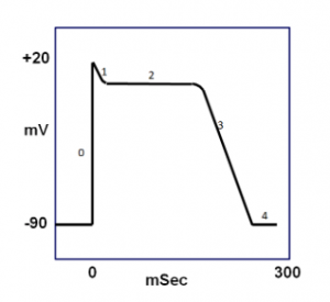
Phase 2 of the diagram indicates which cardiac process?
- Ca2+ channels are open only
- Slow/Fast K+ channels and fast Na+ channels are open only
- Fast Na+ channels are open and Slow/Fast K+ channels are closed
- Slow/Fast K+ channels, Ca2+ channels, and fast Na+ channels are open
- Slow/Fast K+ channels are open onlyAnswer D.
Smooth Muscle Physiology.
-
Which of the following describes the effect of addition of catecholamines, such as norepinephrine and epinephrine, to cardiac muscle cells?
- Increases contractility by increasing Ca2+ influx into the cell
- Increases contractility by decreasing activity of the Na+/K+ ATPase pump
- Decreases contractility by increasing Ca2+ exchange out of the cell
- Decreases contractility by increasing activity of the Na+/K+ ATPase pump
- Catecholamines have no effect on contractility; they only increase transmission of electrical impulses between adjacent cardiac muscle cells.Answer A.
Cardiac Muscle Physiology.
-
A patient with a history of myocardial infarction has _________ preload due to scar tissue in the heart. It requires _________ energy to overcome this.
- Normal, the same
- Decreased, more
- Increased, less
- Increased, more
- Decreased, lessAnswer D.
Cardiac Muscle Physiology.
-
Which step is involved in correcting hypoxia in cardiac muscle?
- Decrease blood flow to lower extremities by vasoconstriction
- Increase production of norepinephrine
- Dephosphorylation of AMP to create adenosine
- Decrease ATP utilization to minimize oxygen consumption in the electron transport chain
- Increase the influx of acetylcholine to stimulate release of sarcoplasmic Ca2+Answer C.
Cardiac Muscle Physiology.
-
Organophosphates are inhibitors of acetylcholinesterase, which leads to an accumulation of acetylcholine. Which effect would we expect to see in a patient that has been exposed to an organophosphate?
- Decreased Ca2+ concentration, tachycardia
- Decreased Ca2+ concentration, bradycardia
- Decreased Ca2+ concentration, normal heart rate
- Increased Ca2+ concentration, tachycardia
- Increased Ca2+ concentration, bradycardiaAnswer B.
Cardiac Muscle Physiology.
-
Which of the following is primarily responsible for maintaining cardiac membrane potential?
- Na+/Ca2+ pump
- Na+/K+ ATPase pump
- ATP exchanger in the sarcoplasmic reticulum
- A & B
- All of the aboveAnswer D.
Cardiac Muscle Physiology.
-
The following best describe which type of muscle: contain sarcomeres, sarcoplasmic reticulum, and T-tubules?
- Skeletal muscle only
- Smooth muscle only
- Cardiac muscle only
- Cardiac and smooth muscle
- Skeletal and smooth muscleAnswer D.
Muscle Physiology.
-
An inhibitor of myosin light chain phosphatase has which effect in smooth muscle?
- Increased sensitivity of myosin and decreased response to calcium
- Increased sensitivity of myosin and increased response to calcium
- Decreased sensitivity of myosin and decreased response to calcium
- Decreased sensitivity of actin and increased response to calcium
- Increased sensitivity of actin and decreased response to calciumAnswer A.
Muscle Physiology.
-
Phase 2 of the diagram indicates which cardiac process?

- Ca2+ channels are open only
- Slow/Fast K+ channels and fast Na+ channels are open only
- Fast Na+ channels are open and Slow/Fast K+ channels are closed
- Slow/Fast K+ channels, Ca2+ channels, and fast Na+ channels are open
- Slow/Fast K+ channels are open onlyAnswer D.
Muscle Physiology.
-
A 19 year old female presents to the emergency room with a serious staph infection covering her abdomen. She explains that she just returned from spring break where became so intoxicated by the pool one day that she puked on herself before passing out in her bikini in the sun. The infection was able to infect her skin via the following means:
- The combination of stomach acid and UVB radiation penetrated the dermis, allowing for infection of the staph bacteria.
- The combination of stomach acid and UVA radiation penetrated the thick skin of the abdomen, allowing for infection of the staph bacteria.
- The spring break party was in Australia where there is a hole in the ozone and the girl was burnt severely from UVC radiation, allowing for infection of the staph bacteria.
- The combination of stomach acid and UVB radiation penetrated the thin skin of the abdomen, allowing for infection of the staph bacteria.
- The combination of UVA and UVB radiation penetrated the thin skin of the abdomen allowing for infection of the staph bacteria.Answer A. Laying in the sun all day would cause severe burning due to the UVB rays. This in combination with the acid from the stomach would cause deeper penetration. Staph bacteria would have to reach the dermis, as this is where the vasculature is to help spread the infection.
Integumentary System Physiology.
-
A skin disorder that results in excessive keratinization charachterized by dryness and fishskin-like scaling:
- Collodionis
- Ichthyosis
- Angiomis
- Nevus flammeus
- AlbanismAnswer B.
Integumentary System Physiology.
-
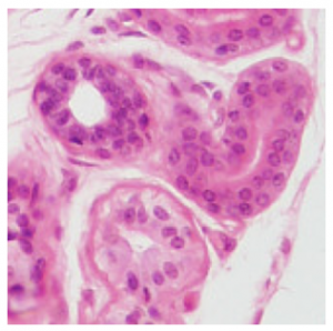
Identify the structure in the image below:

- Arrector pili
- Meissner’s corpuscle
- Eccrine gland
- Sebaceous gland
- Mammary glandAnswer C.
Integumentary System Physiology.
-
All of the following are major features/properties of the skin EXCEPT:
- Production of chemical substances that protect against bacterial growth
- Production of melanin as a defence against UV radiation
- Use of keratin and glycolipids to allow for better absorption of salts
- Secretions that act against bacteria
- DNA electron absorption and conversion of UV radiation to heatAnswer C.
Integumentary System Physiology.
-
Though not always considered an official layer of the skin, the hypodermis is an important structure in our bodies because:
- It provides the primary vasculature to the integument, helping in thermoregulation.
- It stores excess fat so as to avoid excess organ fat causing diseases like fatty liver.
- It is envagenated with ligamentous structures that help hold our organs in place
- It provides a source of energy reserve
- It hosts a number of microscopic structures that aid in course touch sensationAnswer D.
Integumentary System Physiology.
-
The deepest layer of the epidermis, stratum basale, consists of which of the following specialized cells?
- Melanocytes and hair follicles
- Merkel cells and Dendritic cells
- Merkel cells and fibroblasts
- Langerhans cells and subcutaneous adipose tissue
- Adipose tissue and sebaceous glandsAnswer B.
Integumentary System Physiology.
-
Thermoregulation in the skin behaves differently in order to maintain homeostatic needs to the body. With increased temperatures the body will react in which response?
- Increase in body temperature will send signals to the thermoreceptors in the hypothalamus and skin to vasodilate.
- Increase in body temperature will send signals to the thermoreceptors in the thalamus and skin to vasodilate.
- Increase in body temperature will send signals to the thermoreceptors in the hypothalamus and skin to vasoconstrict.
- Increase of body temperature will send signals to the thermoreceptors in the thalamus and skin to vasoconstrict.
- Decrease of body temperatures will send signals to the thermoreceptors in the thalamus and skin to vasoconstrict.Answer A.
Integumentary System Physiology.
-
Which statement is TRUE concerning eccrine and apocrine sweat glands?
- Apocrine sweat glands are smaller than eccrine sweat glands and ducts empty to the surface of the skin.
- Eccrine sweat glands are abundant on palms and soles and are regulated by the parasympathetic nervous system.
- Apocrine sweat glands secrete an odorous substance and eccrine sweat glands empty into hair follicles.
- Eccrine sweat glands secrete a substance that is mostly composed of water and apocrine sweat glands secrete a more viscous substance.Answer D.
Integumentary System Physiology.
-
α-keratin is found in intermediate filaments and expressed in hair, skin, and nails. Which amino acids would most likely enrich Type I α-keratin at a physiological pH to intertwine with Type II α-keratin for the formation of a keratin dimer?
- K,T,Q
- H,G,A
- D,I,H
- G,D,E
- H,K,RAnswer D.
Biochemical Composition of the Skin.
-
Keratin is composed of electrostatic bonds between dimers. The hydrogen bonds and disulfide covalent bonds allow for tetramers to stabilize. The composition of keratin allows for cross linkage to occur in the epidermis through the process of transglutaminase. Which two amino acids must be present for the process to occur in keratinocytes?
- Arginine as the acceptor protein and Glutamine as the donor protein
- Lysine as the donor protein and Arginine as the acceptor protein
- Glutamine as the acceptor protein and Lysine as the donor protein
- Glutamine as the donor protein and Lysine as the acceptor protein
- Arginine as the donor protein and GLutamine as the acceptor proteinAnswer C.
Biochemical Composition of the Skin.
-
UV radiation penetrates the skin damaging the DNA within a keratinocyte. Due to the damage, the body starts a natural reaction for future protection from UV damage. Which answer choice most likely resembles the pathway keratinocytes conduct for further protection?
- The keratinocyte produces POMC via p53 to activate ACTH for anti inflammatory response, thus repairing the damage
- The keratinocyte dies due to p53 activating p21, preventing CDK 1 activation
- The keratinocyte activates p53 which stimulates POMC production generating α-MSH causing pigment production through cAMP cascade
- The keratinocyte signals p53 to produce pigment within the cell via MITF and carry it to the melanocyte via melanosomes
- The keratinocyte is protected from further damage due to p53 causing a POMC production, making 𝝱-endorphins for pigment productionAnswer C.
Biochemical Composition of the Skin.


